Macular Hole
Dedicated to personalized care in a compassionate setting
Macular Hole
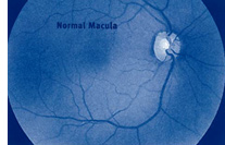 The macula is the central area of one of the most important parts of your eye—the retina.normal The retina is a thin layer of light-sensitive tissue that lines the back of the eye. Light rays are focused onto the retina, where they are transmitted to the brain and interpreted as the images you see. The macula is the portion of the retina responsible for clear, detailed vision.
HOW DOES A MACULAR HOLE FORM?
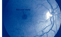 Your eye is filled with a gel-like substance called vitreous, which lies in front of the macula. As you age, the vitreous gel shrinks and pulls away from the macula, usually with no negative effect on your sight. In some cases, however, the vitreous gel adheres to the macula and is unable to pull away. As a result, the macular tissue stretches. After several weeks or months the macula tears, forming a hole. Less common causes of macular holes include injury and long-term swelling of the macula.
WHAT ARE THE SYMPTOMS OF A MACULAR HOLE?
In the early stages of hole formation, your central vision becomes blurred and distorted. If the hole progresses, a blind spot develops in central vision and impairs both distance and near activities.It is important to note that if the macula is damaged, you will not lose your entire vision. You will still have peripheral, or side, vision.
WHAT TESTS WILL BE PERFORMED?
Your ophthalmologist (Eye M.D.) will diagnose a macular hole by looking inside your eye with spedal instruments. To further evaluate the condition of the macula, your ophthalmologist may take special photographs of your eye using a procedure called fluorescein angiography.
HOW IS A MACULAR HOLE TREATED?
Vitrectomy surgery is the most effective treatment to repair a macular hole and possibly improve vision. The surgery involves using tiny instruments to remove the vitreous gel that is pulling on the macula. The eye is then filled with a special gas bubble to help flatten the macular hole and hold it in place while it heals. You must maintain a constant face-down position for one to two weeks after surgery to keep the gas bubble in contact with the macula. A successful result often depends on how well this position is maintained. The bubble will then slowly dissolve on its own. Do not fly in an airplane or travel at high altitudes until the gas bubble has dissolved. A rapid increase in altitude can cause a dangerous rise in eye pressure. You can expect some discomfort after surgery. You will need to wear an eye patch for a short time. Your ophthalmologist will prescribe eyedrops for you and advise you when to resume normal activity. As the macular hole closes, the eye slowly regains part of the lost sight. The visual out come may depend on the size of the hole and how long it was present before surgery. Vision does not return all the way to normal.
WHAT ARE THE RISKS OF VITRECTOMY SURGERY?
Some of the risks of vitrectomy include:
- infection of the eye;
- bleeding of the eye;
- retinal detachment;
- high pressure in the eye;
- poor vision;
- accelerated cataract formation.
It is important that you discuss the potential risks and benefits of this procedure with your ophthalmologist before making a decision regarding treatment.
If you have a Non-urgent matter regarding, Appointment Scheduling, Rescheduling, or Cancellation Please Text (520) 617-2852 for appointment requests.